Pain-induced gamma band oscillations (GBOs) are currently one of the most promising biomarkers of perceived intensity of both stimulus-evoked and spontaneous pain in different species. However, the low spatial resolution of the neuronal population activity sampled from the brain surface and the intrinsic inaccuracy of the source analysis of surface electrocortical data make it difficult to unequivocally identify the neural origin of GBOs. Specifically, whether pain-induced GBOs reflect more stimulus encoding in the primary somatosensory cortex (S1) or result from motor-related activity for nocifensive behavior in the primary motor cortex (M1) is much debated. Recently, Dr. Lupeng Yue and Professor Li Hu from the Institute of Psychology, Chinese Academy of Sciences, recorded neural activity simultaneously from the brain surface and from different depths of the bilateral S1/M1 in freely-moving rats receiving nociceptive stimulation. This approach enables scientists to assess the relationship of neural responses at different spatial scales. Specifically, the possibility of recording pain-induced GBOs at the mesoscopic scale of local field potentials (LFPs) allows bridging the gap between the macroscopic scale of electroencephalogram/electrocorticogram (EEG/ECoG) and the microscopic scale of single-unit spiking. By comprehensively exploring the contribution of superficial vs deep cortical layers of bilateral S1 and M1 in the generation of pain-induced GBOs, the authors found that GBOs measured from superficial layers of S1 contralateral to the stimulated paw not only had the largest magnitude, but were also showed the strongest coupling with epidural GBOs in terms of both temporal sequence and phase consistency (Figure 1). In addition, spiking of interneurons in superficial S1 layers had the strongest coherence with the phase of epidural GBOs (Figure 2). This set of results provide the first direct demonstration that pain-induced GBOs measured at population level, the most promising biomarker of pain perception so far, reflect neural activity coupled with the spike firing of interneurons located in the superficial layers of S1 contralateral to the side of nociceptive stimulation. This discovery has major clinical significance, as a better characterization of the functional significance of pain-induced GBOs will increase their efficacy in the diagnosis and prediction of treatment outcome in various pain conditions. This work entitled “The Neural Origin of Nociceptive-Induced Gamma-Band Oscillations” was published in the Journal of Neuroscience on April 22. It was funded by the National Natural Science Foundation of China (Grants 31822025 and 31671141) and the Scientific Foundation Project of Institute of Psychology, Chinese Academy of Sciences (Grant Y6CX021008). 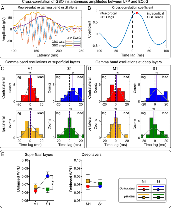
Figure 1 The temporal sequence and phase relationship between GBOs measured intracortically and epidurally. 
Figure 2 The relationship of spike firing in bilateral S1 and M1 and the laser-induced GBOs simultaneously recorded from the brain surface. CONTACT:
Ms.LIU Chen
Institute of Psychology, Chinese Academy of Sciences
Beijing 100101, China.
E-mail: liuc@psych.ac.cn |